
NML Research Article | Targeted detection and treatment of tumors in vivo: dual-modal PET/optical imaging system based on SiO2 hollow core-shell nanostructures
In Vivo Tumor-Targeted Dual-Modality PET/Optical Imaging with a Yolk/Shell-Structured Silica Nanosystem
Sixiang Shi, Feng Chen, Shreya Goel, Stephen A. Graves, Haiming Luo, Charles P. Theuer, Jonathan W. Engle, Weibo Cai
Nano-Micro Lett. (2018) 10:65
https://doi.org/10.1007/s40820-018-0216-2
Highlights of this article
1 Using quantum dots as the core and mesoporous SiO2 hollow spheres as the shell, a hollow core-shell structured nano-system was prepared.
2 Combining the advantages of PET and optical imaging, use dual-modal PET/optical imaging for collaborative cancer diagnosis.
3 Successfully realized the targeted therapy of tumor blood vessels, increasing the residence time and target specificity of the drug at the tumor site.
brief introduction
PET (Positron emission tomography, positron emission tomography) is the most advanced clinical examination imaging technology in the field of nuclear medicine. The advantages of high resolution enable it to be widely used in clinical diagnosis of tumors and heart and lung diseases. Bimodal or multimodal molecular probe technology can obtain more comprehensive information in diagnosis or treatment.
In the field of biomedicine, mesoporous silicon materials have the characteristics of large specific surface area, large pore volume and diverse pore structure, and have good application prospects in drug transportation. The key to realizing drug delivery is that mesoporous silicon can effectively encapsulate different types of drug molecules in the mesoporous silicon pores, thereby preventing the therapeutic drugs from enzymatic hydrolysis.
The Weibo Cai team at the University of Wisconsin in the United States designed a hollow core-shell structured SiO2 nanosystem for tumor blood vessel targeting and bimodal PET/optical imaging: 64Cu-NOTA-QD@HMSN-PEG-TRC105.
The system has good targeting specificity and excellent imaging capabilities, making the diagnosis results more reliable. This work combines vasculature targeting, pH-sensitive drug delivery, and dual-modality imaging into a single platform. The hollow core-shell structured SiO2 nanosystem is applied to image-guided tumor-targeted drug delivery, effectively enhancing cancer Diagnosis and treatment.
Graphic guide
1 Synthesis of QD@HMSN
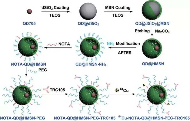
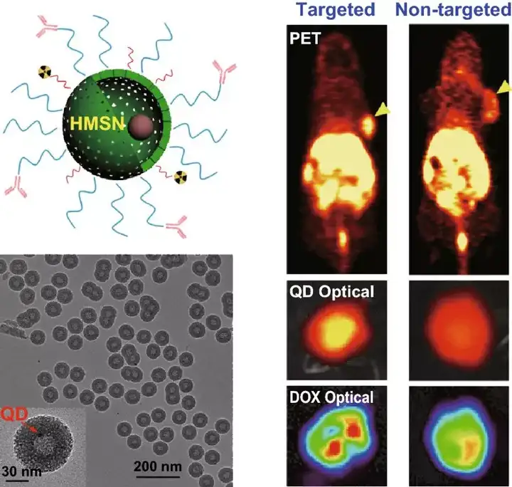
Schematic diagram of synthesis and functionalization of QD@HMSN SiO2 hollow core-shell nanostructures.
2 In vivo blood vessel targeting and PET imaging
2 In vivo blood vessel targeting and PET imaging
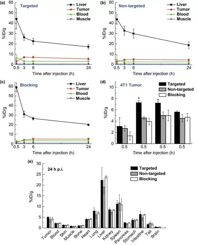
Quantitative data obtained from PET images and ROI analysis showed that the tumor uptake in the targeted group was significantly enhanced compared with the non-targeted group and the blocked group.
3 Drug loading and optical imaging
3 Drug loading and optical imaging
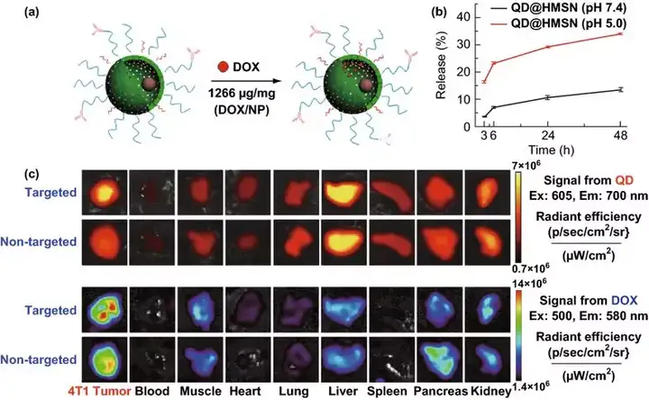
In QD@HMSN NPs with hollow core-shell nanostructures, the drug loading increased significantly to 1266 mg/g (DOX weight/NP weight).
The increased drug load may be beneficial to enhance the curative effect of cancer chemotherapy, and at the same time, the nanoparticle dose required for each treatment is smaller, thereby reducing the in vivo cytotoxicity of the nanocarrier.
The increased drug load may be beneficial to enhance the curative effect of cancer chemotherapy, and at the same time, the nanoparticle dose required for each treatment is smaller, thereby reducing the in vivo cytotoxicity of the nanocarrier.

About the Author

Main research directions:
1) Development of multi-modal molecular imaging agents; 2) Molecular therapy of cancer; 3) Nanotechnology and its biomedical applications.
Research group homepage:
http://mi.wisc.edu/index.htm
This information is sourced from the Internet for academic exchanges only. If there is any infringement, please contact us to delete it immediately.
18915694570
Previous: Soochow University & U